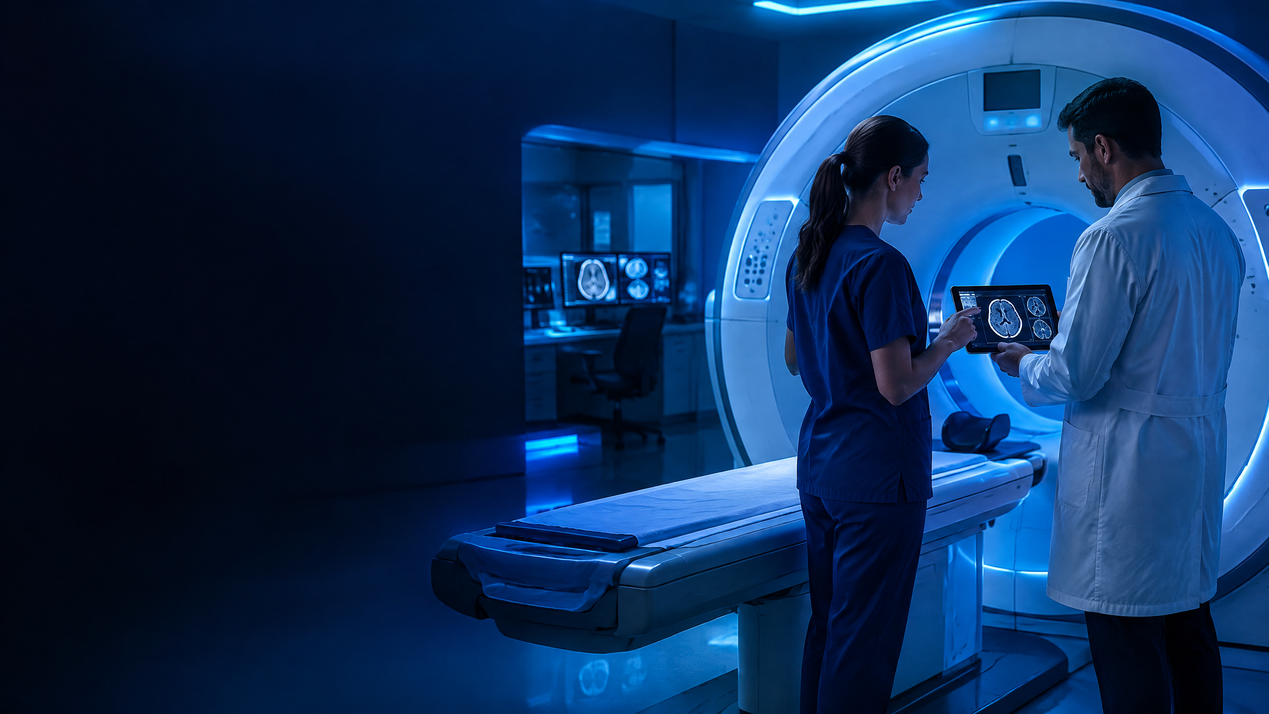
Enter the exam experience before clinical practice.
Atlas Imaging Lab is the visual learning companion for Atlas Método training and postgraduate courses. Students navigate CT, MRI and radiography protocols, connect slices to anatomy and turn class content into deliberate practice.
Try the multiplanar logic
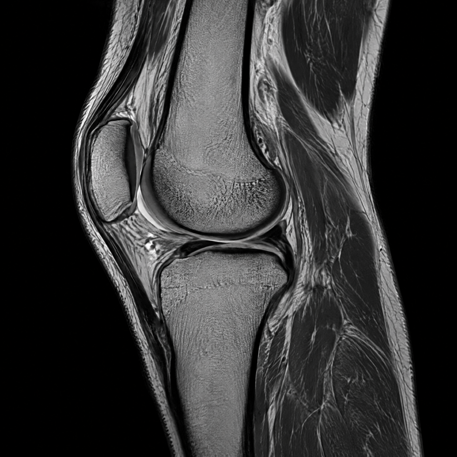
Switch between a head CT and knee MRI, move the teaching position, and observe the relationship among axial, coronal, and sagittal planes.
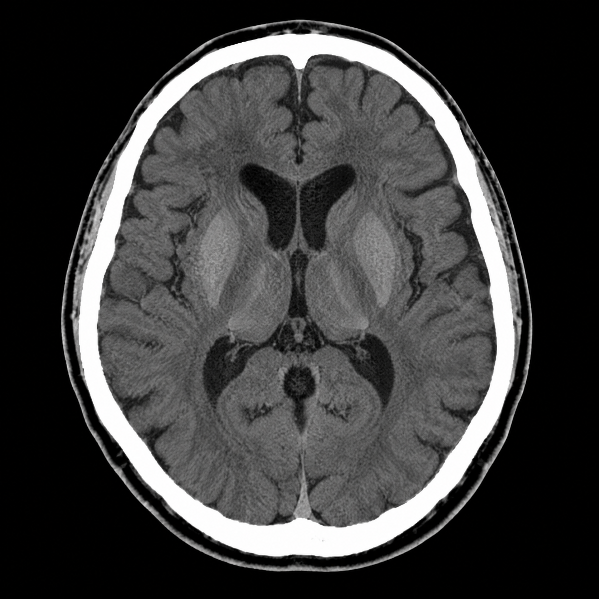
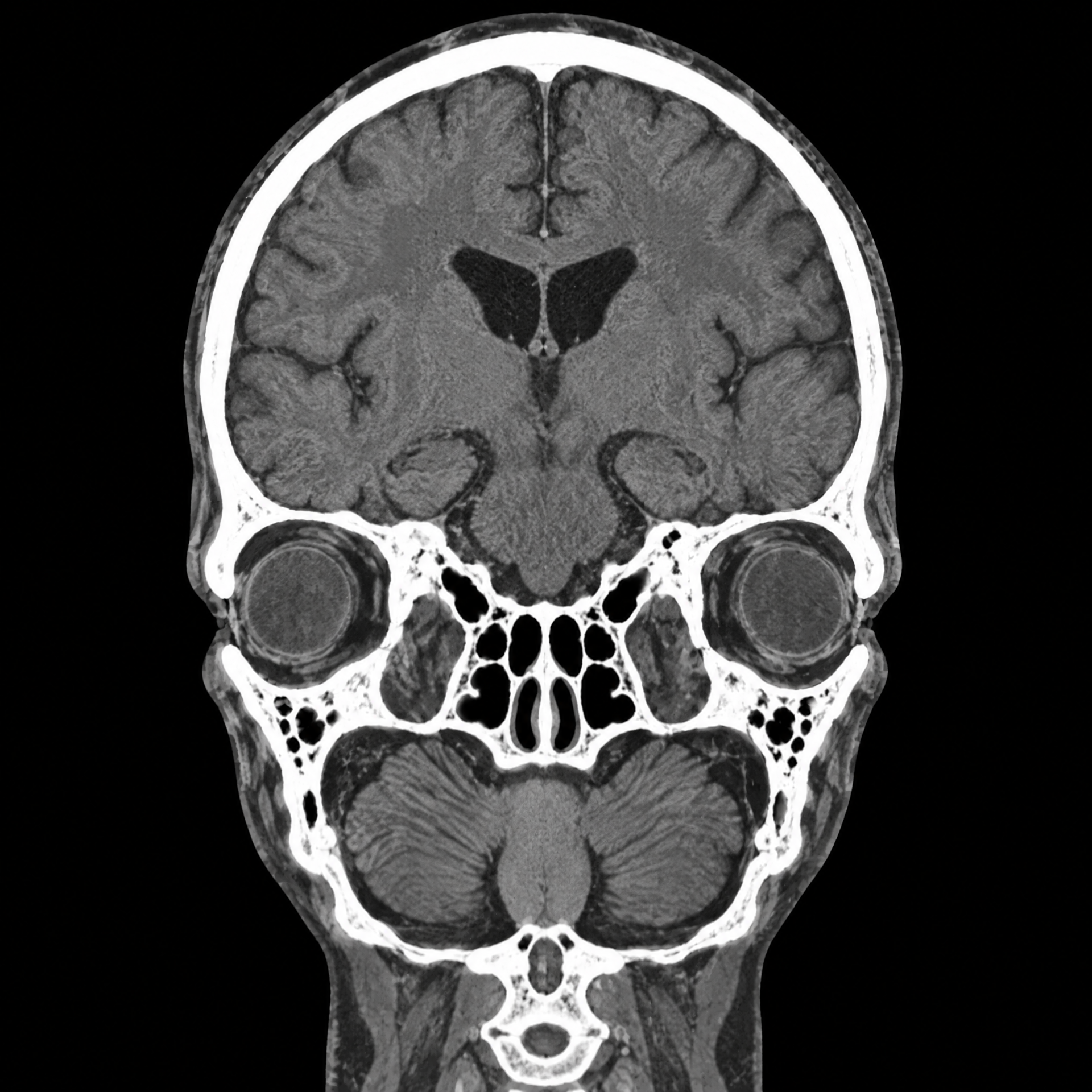
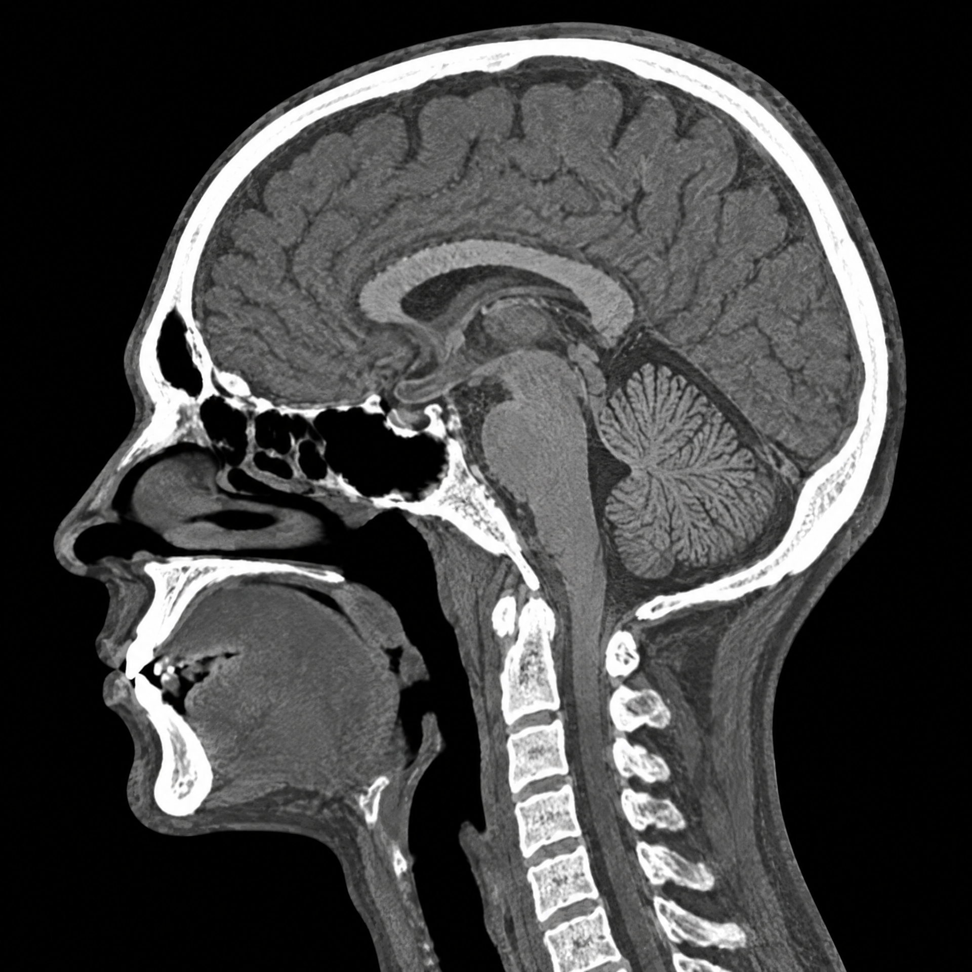
Limited preview with synthetic images and a synchronized cursor. The full lab uses approved series, windowing controls, Stack/MPR, and DICOM gates.
Before class
Review anatomy, planes and protocol goals to arrive with a mental map.
During the course
Follow slices, series and windows while the instructor discusses technique and anatomic recognition.
After class
Repeat navigation, use the 3D Atlas, take the quiz and track progress by protocol.
From the classroom to image-based reasoning.
The lab organizes the experience into steps so learners know what to observe, how to orient themselves and where to review.
Learn what changes among CT, MRI and radiography.
Each modality requires a different orientation strategy. Content does not simulate diagnosis: it teaches anatomy, acquisition, presentation and systematic review.
TCComputed tomography
Navigate series, correlate axial, coronal and sagittal planes, and understand windows, reconstruction and contrast phases.
 RM
RMMagnetic resonance imaging
Compare sequences and planes using Stack or MPR only when geometry is compatible.
 RX
RXRadiography
Recognize projection, positioning, technical quality and two-dimensional anatomy in dedicated paths.
The exam becomes a learning path, not just an image sequence.
Select the protocol, understand the series, navigate the planes and consolidate. Anatomy, exam and feedback remain in one journey.
Explore the studiesChoose protocol and modality
Body region, CT/MRI/X-ray, difficulty and learning goals.
Understand series and acquisition
Plane, phase, reconstruction, sequence and window without mixing concepts.
Navigate and correlate
Viewer-contained scrolling, synchronized planes and Anatomy × Exam.
Review and track progress
Contextual quiz, formative explanations and a session report.
More than a viewer: a learning workstation.
The Viewer is the core, connected to the 3D Atlas, library, annotations, learning files and live classes.
DICOM laboratory
Protected Stack/MPR, window controls, measurements, A/B comparison and automatic recovery.
Open libraryContextual 3D Atlas
Structures, anatomy function, protocol correlation and Portuguese videos by region.
Open 3D AtlasImage-based quizzes
Protocol-linked questions, visual references, explanations and shuffled new rounds.
Explore quizzesLearning base
Books, articles, live classes and editorial materials organized around the course.
View learning files
Study the exam as it is discussed in class.
Sign in to access approved series and the full lab. The experience is educational and does not replace supervision, institutional protocols or diagnostic interpretation.