The interface is translated. Medical content remains in its editorial source language until specialist review.
Expanded visual content

Radiology library

Browse body regions, modalities, levels and anatomy points. Each module combines a teaching series, annotations and exercises.

14 published studies1444 teaching images8 indexed regions

Mapa de estudo

1

indexed regions

anatomia e sistemas

8
2

Modality

TC, RM, RX e outros

3
3

published studies

protocolos navegáveis

14
Try the atlas

3D spatial index

Explore the body by region

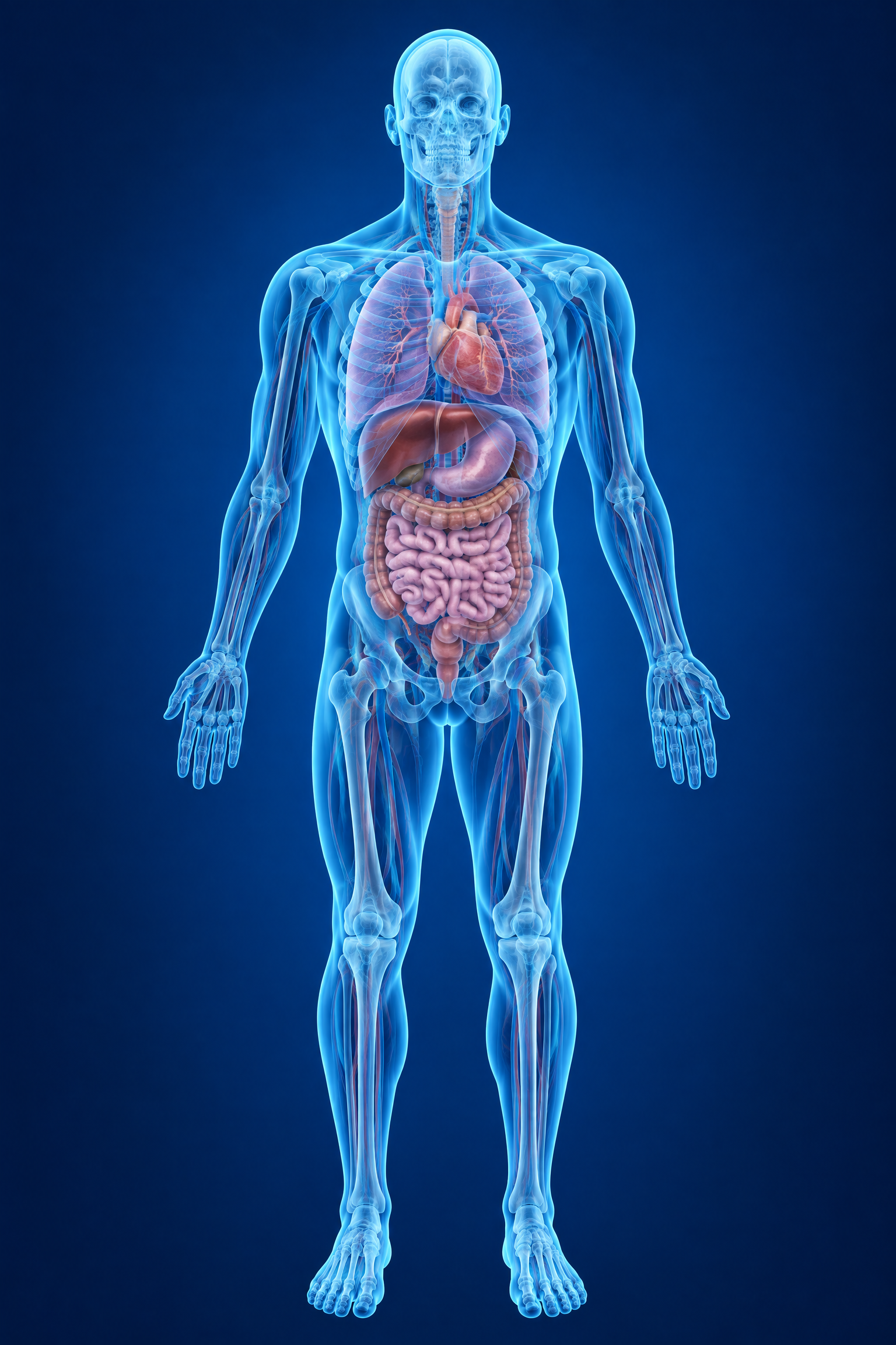
Select a region directly on the anatomical render or use the accessible list.

Anterior view
3D anatomical render, Anterior view
AvailableSelected

Current selection

All regions are visible in the library.

Active filters

14 studies found

Head and brainFace and neckSpineChestAbdomenPelvis and hipUpper limbsLower limbs
Use the blue points on images to review landmarks.
Study image: Basic radiography: anatomy and technique
RXFeaturedEditorial translation under review
Multiple regions
18 mins6 imagesBeginner

Basic radiography: anatomy and technique

Introductory track with open projections of the chest, knee, shoulder, pelvis, hand and elbow to study projected anatomy, orientation and physical principles.

X-raysProjectionsBone anatomy
Open study
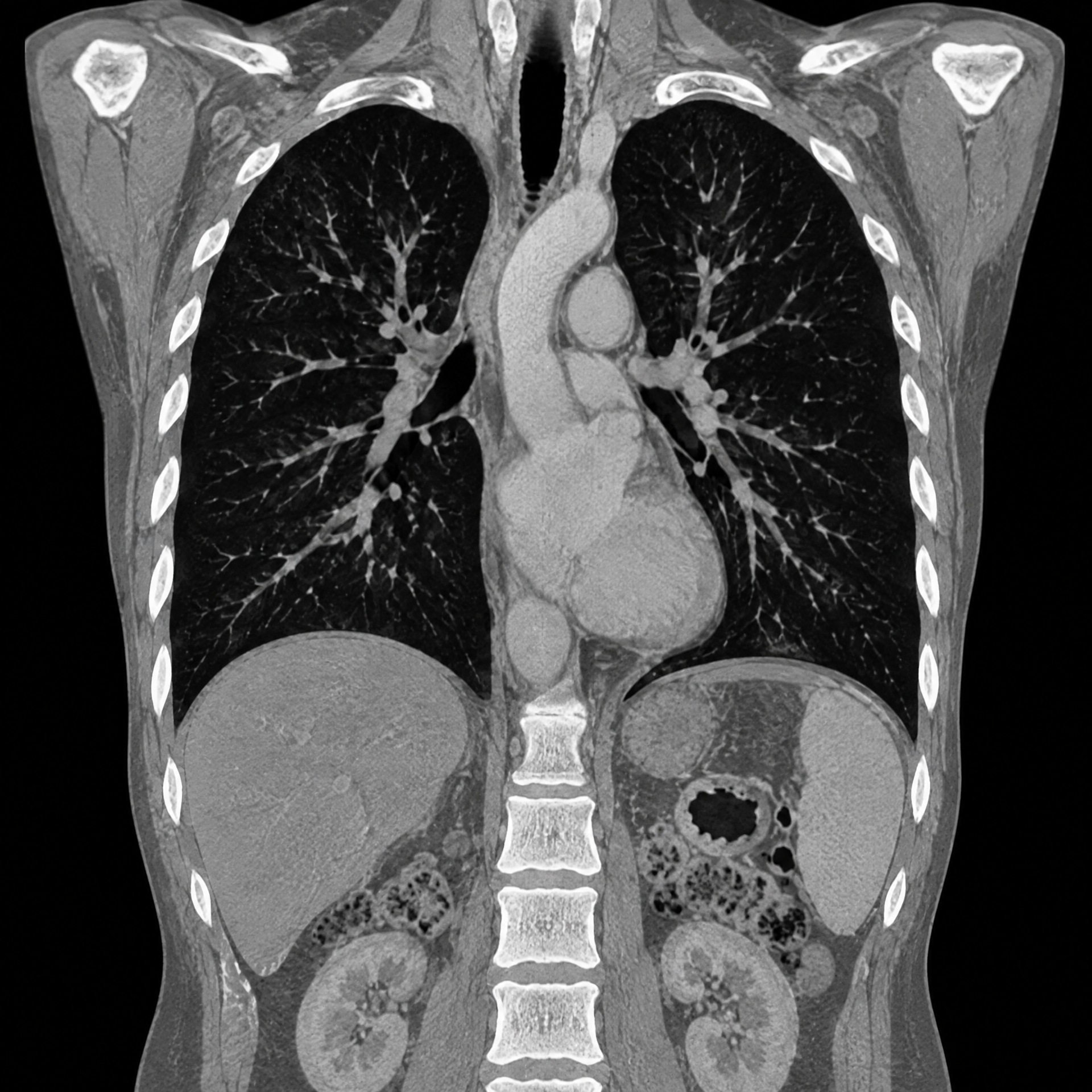
Study image: Thoracic anatomy on CT
TCFeaturedEditorial translation under review
Thorax
12 mins554 imagesBeginner

Thoracic anatomy on CT

Visual review of the lungs, mediastinum and bony thorax with a didactic series for orientation by level.

LungsMediastinumMultiplanar
Open study
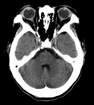
Study image: CT of the cranium
TCEditorial translation under review
Cranium and brain
10 mins423 imagesBeginner

CT of the cranium

Routine non-contrast CT of the cranium

craniumbrain
Open study
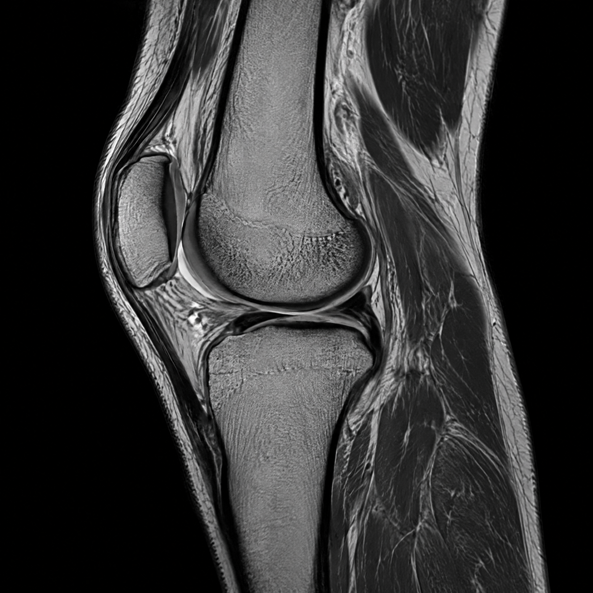
Study image: Multiplanar knee MRI
RMEditorial translation under review
Lower extremities
14 mins136 imagesAdvanced

Multiplanar knee MRI

Introduction to systematic interpretation of the knee magnetic resonance imaging (MRI) and its main osteoarticular landmarks.

KneeOsteoarticularMultiplanar
Open study
No thumbnail
TCEditorial translation under review
Lower extremity
10 mins307 imagesBeginner

Knee CT

Knee structure analysis

KneeLower extremitySkeletal
Open study
Study image: Ankle and foot radiography — alignment and trauma
RXEditorial translation under review
Lower limbs
15 mins4 imagesBeginner

Ankle and foot radiography — alignment and trauma

Didactic draft of ankle and foot radiography — alignment and trauma, with planned series/projections and linked anatomic repertoire. Publication conditional on editorial review and eligible images.

X-rayAnkle and footP1 in trauma and bone pain; weight-bearing is selective [40] [41]
Open study
No thumbnail
TCEditorial translation under review
Lower limbs
15 mins0 imagesIntermediate

CT of the ankle/foot — complex osseous trauma

Didactic draft of CT of the ankle/foot — complex osseous trauma, with planned series/projections and linked anatomic repertoire. Publication conditional on editorial review and eligible images.

CTAnkle and footP1 when the question is complex fracture, calcaneus, talus, fragment or planning [40] [42]
Open study
Study image: Knee X-ray — trauma, alignment and selective weight-bearing
RXEditorial translation under review
Lower limbs
15 mins2 imagesBeginner

Knee X-ray — trauma, alignment and selective weight-bearing

Didactic draft of knee radiography — trauma, alignment and selective weight-bearing, with planned series/projections and linked anatomic repertoire. Publication conditional on editorial review and eligible images.

X-rayKneeP1 in selected trauma, chronic pain and alignment; weight-bearing/tunnel are complementary [35] [36]
Open study
Study image: Pelvis and hip radiography — trauma basics and alignment
RXEditorial translation under review
Pelvis and hip
15 mins1 imagesBeginner

Pelvis and hip radiography — trauma basics and alignment

Didactic draft of pelvis and hip radiography — trauma basics and alignment, with planned series/projections and linked anatomic repertoire. Publication is conditional on editorial review and eligible image linkage.

RadiographyPelvis and hipP1 for trauma, fracture, dislocation, alignment and bone pain; frog‑leg is avoided when there is suspicion of proximal fracture [19] [20]
Open study
Study image: Chest radiography PA and lateral — global anatomy
RXEditorial translation under review
Thorax
15 mins2 imagesBeginner

Chest radiography PA and lateral — global anatomy

Didactic draft of chest radiography PA and lateral — global anatomy, with planned series/projections and linked anatomic repertoire. Publication conditioned on editorial review and eligible images.

RadiographyThoraxP1 for global overview and initial correlation; portable AP is a conditional alternative and does not equate to PA [13] [14]
Open study
Study image: Elbow X-ray — bone, alignment and effusion
RXEditorial translation under review
Upper extremities
15 mins4 imagesBeginner

Elbow X-ray — bone, alignment and effusion

Educational draft of an elbow X-ray — bone, alignment and effusion, with planned series/projections and linked anatomic repertoire. Publication conditional on editorial review and linkage to eligible images.

X-rayElbowP1 as initial exam in acute and chronic pain; CT and MRI are complementary [25] [26]
Open study
Study image: Radiography of the hand — PA, oblique and fan lateral
RXEditorial translation under review
Upper extremities
15 mins2 imagesBeginner

Radiography of the hand — PA, oblique and fan lateral

Didactic draft of hand radiography — PA, oblique and fan lateral, with planned series/projections and linked anatomic repertoire. Publication conditional on editorial review and linkage to eligible images.

RadiographyHandP1 for trauma and bone [31] [32]
Open study
Study image: Shoulder X-ray — initial and trauma series
RXEditorial translation under review
Upper limbs
15 mins1 imagesBeginner

Shoulder X-ray — initial and trauma series

Didactic draft of shoulder radiography — initial and trauma series, with planned series/projections and linked anatomic repertoire. Publication conditioned on editorial review and eligible images.

X-rayShoulderP1 for trauma, fracture, dislocation and initial bony assessment [22] [23]
Open study
Study image: Wrist X-ray — initial series and carpal alignment
RXEditorial translation under review
Upper limbs
15 mins2 imagesBeginner

Wrist X-ray — initial series and carpal alignment

Didactic draft of wrist radiographs — initial series and carpal alignment, with planned series/projections and linked anatomic repertoire. Publication conditional on editorial review and eligible images.

X-rayWristP1 for fracture, alignment and instability; targeted projections are additional [28] [29]
Open study